News - NeuroAnalytica is now released with a free license for nonprofit and academic research.
Email info@neuroanalytica.com to register and receive download information (64-bit linux only). Please include your name, institution, department, and how you plan to use NeuroAnalytica.
Please visit the resource pages for installation and usage information.
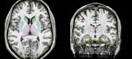
The methods used by Brain Image Analysis in NeuroAnalytica are a commercial extension of BRAINS and AutoWorkup (published January, 2011), the result of over 20 years of brain research and software development. Our results are very stable across different scanners, scanner vendors and console upgrades. Perfect for multi-site studies, as shown in the AutoWorkup paper.
Clinical Trials
We are continuing to develop NeuroAnalytica be 21 CFR compliant as a medical device, and will soon be announcing a number of developments in this area. A full battery of highly reliable, validated brain measures will be available. Please contact us if you are interested in any further information about clinical applications.
Tweet